Development of Tooth
The development of the tooth involves many complex biological processes, including epithelial mesenchymal interactions, morphogenesis and mineralization.
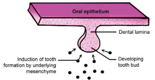
In human beings, 20 deciduous and 32 permanent teeth develop from the interaction between the oral epithelium cells and the underlying mesenchymal cells.
Primary Epithelial Band
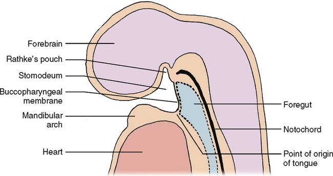
The oral ectoderm is neural crest or ectomesenchyme in origin. It is lined by stratified squamous epithelium.The initial oral cavity develops after the rupture of the buccopharyngeal membrane at the fourth week of intrauterine life.
 |
| Buccopharyngeal membrane |
The interaction between the oral epithelium and the underlying mesenchymal cells results
in tooth development.
After thirty-seven (37 days or 5th week) days of development, a continuous band of thickened epithelium forms around the mouth in both the future upper and lower jaws.
 |
| Primary Epithelium Band |
These bands of the epithelium are roughly horseshoe-shaped structures.
Dental Lamina
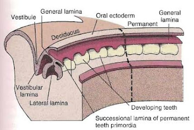
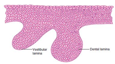
At the sixth week of gestation period,The primary epithelial band forms two subdivisions called the dental lamina and the vestibular lamina.
 |
| Dental Lamina |
The dental lamina is a band of epithelium that has invaded the underlying ectomesenchyme along both the horseshoe-shaped future dental arches.
 |
| Dental Lamina |
Deciduous dentition develops directly from the dental lamina at the eighth week of fetal life, whereas the permanent molars develop from a distal extension of the dental lamina.
 |
| Dental Lamina |
The lingual extension of dental lamina is called successional lamina. Successional lamina is responsible for the development of permanent incisors, canine and premolars.
Fate of Dental Lamina
After initiation of tooth development, the dental lamina degenerates. Total functional activity period of the dental lamina is around five years.
The remnants of the dental lamina may persist in the jaw or gingiva in the form of islands or epithelial pearls. These are known as cell rest of Serres.
Vestibular Lamina
 |
| Vestibular Lamina |
Facial (labial and buccal) to dental lamina another thick band of epithelium develops in the maxillary and mandibular dental arches. It is called as the vestibular lamina or the lip furrow band.
Tooth Development Anomalies
Anodontia and Oligodontia
Anodontia is the congenital absence of tooth germ of the entire dentition. Absence of single tooth germ or multiple tooth germs is termed oligodontia. The teeth commonly found missing are upper lateral incisors, third molars and lower second premolars.
Supernumerary Teeth
Teeth which are present in addition to the normal number of teeth are called supernumerary teeth. This occurs due to hyperactivity of the dental lamina, leading to the initiation of additional tooth buds.
The most common supernumerary teeth are the mesiodens which are present between two upper central incisors and paramolars present by the side of the molars.
Tooth Development Charts
Stages of Tooth Development (On the basis of Shape of Enamel Organ)
The stages of tooth development can be divided into the following stages on the basis of the shape of the enamel organ:
1. Bud stage
2. Cap stage
3. Bell stage- (a) Early Histodifferentiation (b) Advanced Morphodifferentiation
4. Advanced bell stage
The Histology of each Tooth Development Stages is well explained with Diagrams.
The Histology of each Tooth Development Stages is well explained with Diagrams.
Bud Stage (Initiation)
Enamel Organ
The enamel organ, which looks like a bud, consists of low columnar cells in the periphery and polygonal cells in the center.
Increased proliferation of ectomesenchymal cells and migration of neural crest cells, the cellular density of ectomesenchyme immediately adjacent to the enamel organ increases. This is referred to as condensation of ectomesenchyme.
 |
| Bud Stage |
Increased proliferation of ectomesenchymal cells and migration of neural crest cells, the cellular density of ectomesenchyme immediately adjacent to the enamel organ increases. This is referred to as condensation of ectomesenchyme.
Dental Papilla
The condensed ectomesenchyme immediately under the enamel organ is the dental papilla.
Dental Follicle
The condensed ectomesenchyme surrounding the enamel organ and the dental papilla is the dental follicle/sac.
Tooth Development Anomalies
Macrodontia and Microdontia
Macrodontia is an abnormally larger tooth and microdontia is an abnormally smaller tooth. This can occur because of abnormal proliferation of the tooth germ at the bud stage and can affect a single tooth or the complete dentition.
Cap Stage (Proliferation)
Enamel Organ
The enamel organ shows an unequal rate of proliferation in different parts instead of uniform expansion. This leads to a stage where the enamel organ looks like a cap.
 |
| Cap Stage |
The cells of the enamel organ in the convex portion of the cap are cuboidal in shape and
form the outer enamel epithelium.
The cells in the concavity of the cap are columnar in shape and form the inner enamel epithelium.
The outer enamel epithelium is separated from the dental sac and the inner enamel epithelium is separated from the dental papilla by a basement membrane.
Polygonal cells present in the center of the enamel organ between the inner and the outer enamel epithelium are Stellate Reticulum.
The proteinaceous fluid-containing albumin gives a cushion-like consistency to the stellate reticulum that supports and protects the delicate enamel-forming cells.
 |
| Cap Stage |
Transient Structures in Enamel Organ
Primary Enamel Knot The cells in the centre of the concavity of the ‘cap’ of the enamel organ are densely packed and form a knob-like enlargement projecting towards the underlying dental papilla.
Enamel Cord vertical extension of the enamel knot running across the centre of the enamel organ.
Enamel Septum when this extension meets the outer enamel epithelium.
Enamel Navel A small depression that forms at the junction between the enamel septum and the outer enamel epithelium.
Enamel Niche During sectioning of the developing tooth tissue specimen, the enamel organ appears to be attached to the oral epithelium by two or more strands of dental lamina. This artefactual stalk is called enamel niche.
Dental Papilla
Cells of the dental papilla appear more crowded in this stage. The dental papilla also shows signs of becoming more vascular. This is evident by the presence of mitotic figures and active budding of capillaries.
Dental Sac
The dental sac appears more condensed and fibrous.
Tooth Development Anomalies
Gemination and Fusion
Gemination and Fusion
Gemination is division of tooth germ while fusion is the union of the adjacent tooth germs which can occur during the cap stage of tooth development.
Dens Invaginatus
Abnormal invagination of the enamel organ into the dental papilla can cause an appearance of tooth within the tooth called dens invaginatus or dens in dente. This abnormality appears as a deep lingual pit which may lead to pulp exposure warranting endodontic therapy of the affected tooth.
Bell Stage (Histo and Morpho Differentiation)
Enamel Organ
In this stage, the crown of the tooth gets its final shape (morphodifferentiation) and the cells that form the hard tissues of the crown (the ameloblasts that form the enamel and the odontoblasts that form dentin) acquire histodifferentiation.
Four different types of epithelial cells of the enamel organ can be identified in this stage:
A. Inner enamel epithelium
B. Stratum intermedium
C. Stellate reticulum
D. Outer enamel epithelium
The junction between the inner and outer enamel epithelia is known as the zone of reflexion
or cervical loop.
A. Inner Enamel Epithelium
This single layer of columnar cells differentiates into tall columnar cells called ameloblasts before the formation of enamel (amelogenesis).
B. Stratum Intermedium
Cells seen between the inner enamel epithelium and stellate reticulum are called stratum intermedium. They are associated with protein synthesis and transport of nutrients to the ameloblasts. The role of stratum intermedium is to regulate the formation of enamel.
C. Stellate Reticulum
These star-shaped cells are connected to one another and to the cells of the outer enamel epithelium and the stratum intermedium by desmosomes.
The role of stellate reticulum is to protect the underlying inner enamel epithelial cells. After a layer of dentin is laid down, the inner enamel epithelial cells are deprived of their nutritional supply from the dental papilla.
The stellate reticulum collapses before the formation of enamel, bringing the capillaries in the dental follicle closer to the inner enamel epithelial cells to provide nutrition.
D. Outer Enamel Epithelium
The outer enamel epithelium has cuboidal cells. They are connected to the adjacent cells by junctional complexes.
Dental Papilla
Fine fibrils from the basal lamina, which separates the dental papilla from the enamel organ,
extend into an acellular zone. The acellular zone is present between the basal lamina and the dental papilla and is devoid of cells.
The peripherally placed undifferentiated ectomesenchymal cells of the dental papilla increase in size before the enamel formation begins. They are initially cuboidal and later become columnar and differentiate into odontoblasts.
In the absence of the inner enamel epithelial cells, differentiation of odontoblasts does not occur and dentin does not form.
The basement membrane which separates the enamel organ and dental papilla just before dentin formation is called membrana preformativa.
Dental Follicle
As more and more of collagen fibrils occupy the extracellular spaces between the fibroblasts of the dental follicle, these can be clearly distinguished from the more vascular dental papillae.
It is these fibres of the dental follicle that later differentiate into periodontal fibres that become embedded in the cementum and alveolar bone.
Tooth Development Anomalies
Dens Evaginatus
It is cusp-like elevation seen in the occlusal surface of premolars or molars. This aberrancy occurs due to the abnormal proliferation of inner enamel epithelium into the stellate reticulum resulting in a core of dentin with intervening pulpal tissue covered by enamel.
With occlusal wear or fracture of this cusp-like structure, pulp exposure and pulpal necrosis can occur warranting endodontic therapy.
 |
| Bell Stage |
A. Inner enamel epithelium
B. Stratum intermedium
C. Stellate reticulum
D. Outer enamel epithelium
The junction between the inner and outer enamel epithelia is known as the zone of reflexion
or cervical loop.
A. Inner Enamel Epithelium
This single layer of columnar cells differentiates into tall columnar cells called ameloblasts before the formation of enamel (amelogenesis).
B. Stratum Intermedium
Cells seen between the inner enamel epithelium and stellate reticulum are called stratum intermedium. They are associated with protein synthesis and transport of nutrients to the ameloblasts. The role of stratum intermedium is to regulate the formation of enamel.
C. Stellate Reticulum
These star-shaped cells are connected to one another and to the cells of the outer enamel epithelium and the stratum intermedium by desmosomes.
The role of stellate reticulum is to protect the underlying inner enamel epithelial cells. After a layer of dentin is laid down, the inner enamel epithelial cells are deprived of their nutritional supply from the dental papilla.
The stellate reticulum collapses before the formation of enamel, bringing the capillaries in the dental follicle closer to the inner enamel epithelial cells to provide nutrition.
D. Outer Enamel Epithelium
The outer enamel epithelium has cuboidal cells. They are connected to the adjacent cells by junctional complexes.
 |
| Bell Stage |
Dental Papilla
Fine fibrils from the basal lamina, which separates the dental papilla from the enamel organ,
extend into an acellular zone. The acellular zone is present between the basal lamina and the dental papilla and is devoid of cells.
The peripherally placed undifferentiated ectomesenchymal cells of the dental papilla increase in size before the enamel formation begins. They are initially cuboidal and later become columnar and differentiate into odontoblasts.
In the absence of the inner enamel epithelial cells, differentiation of odontoblasts does not occur and dentin does not form.
The basement membrane which separates the enamel organ and dental papilla just before dentin formation is called membrana preformativa.
Dental Follicle
As more and more of collagen fibrils occupy the extracellular spaces between the fibroblasts of the dental follicle, these can be clearly distinguished from the more vascular dental papillae.
It is these fibres of the dental follicle that later differentiate into periodontal fibres that become embedded in the cementum and alveolar bone.
Tooth Development Anomalies
Dens Evaginatus
It is cusp-like elevation seen in the occlusal surface of premolars or molars. This aberrancy occurs due to the abnormal proliferation of inner enamel epithelium into the stellate reticulum resulting in a core of dentin with intervening pulpal tissue covered by enamel.
With occlusal wear or fracture of this cusp-like structure, pulp exposure and pulpal necrosis can occur warranting endodontic therapy.
Advanced Bell Stage
Advanced bell stage is characterized by the beginning of mineralization and root formation.
 |
| Advanced Bell Stage |
Mineralization
The line separating the newly differentiated odontoblasts and the inner enamel epithelial cells outlines the dentinoenamel junction.
The first layer of dentin is formed along the future dentinoenamel junction and formation proceeds pulpally and apically. After the formation of first layer of dentin, enamel is laid down over the dentin by the ameloblast and enamel formation proceeds occlusally.
Tooth Development Anomalies
Tetracycline Staining
Tetracycline Staining
Tetracycline is an antibiotic which has high affinity for calcified tissue. Ingestion of this antibiotic during the mineralization of enamel and dentin can lead to discoloration. This consequent discoloration of enamel and dentin occurring at the time of tooth development is called tetracycline staining.
Crown Pattern Determination
 |
| Crown Pattern Determination |
Transition of the enamel organ from cap to bell stage was because of the invagination of the inner layer of the enamel organ and the growth of the enamel organ at its margins.
Earlier, it was thought that the infolding of the enamel organ occurs as a result of the pressure exerted by the proliferating cells of the dental papilla on the inner enamel epithelium. Now, studies have shown that this pressure is opposed equally by the fluid present in the stellate reticulum.
The infolding which gives shape to the crown occurs as a result of differences rates of mitotic division and differentiation process of cells of the inner enamel epithelium.
Those inner enamel epithelial cells which lie in the future cusp tip region stop dividing earlier and begin to differentiate first to assume their function of producing enamel. This point at which differentiation of inner enamel epithelial cells occurs first is known as the growth center
The other cells of the enamel organ continue to divide, exerting pressure on the cells in the cusp tip region. This causes the cells in the region of cusp tip to be pushed up into the enamel organ towards the outer enamel epithelium.
This process determines the shape of the area between the two cusp tips that is the extent of cuspal slopes and the cusp height.
Development of Root
The
enamel and dentin formation reaches the future cementoenamel junction, root
development begins From the cervical portion of the enamel organ arises Hertwig’s
epithelial root sheath (HERS).
 |
| Development of Root |
Hertwig’s epithelial root sheath is double layered. It contains only the outer and the inner enamel epithelia and not the stratum intermedium and stellate reticulum.
Enamel
on radicular portion is not formed because of the absence of stratum
intermedium.
The
root sheath initiates formation of dental root and determines the number,
shape, length and dimensions
of the roots.
After
the first layer of radicular dentin is formed, the HERS loses its continuity
and at the same time the cells of the dental follicle/sac proliferate and invade
the double layer of HERS, dividing it into epithelial strands.
This
results in differentiation of cementoblasts from the cells of the dental follicle,
which deposit cementum on the surface of the radicular dentin. Similarly,
fibroblasts and osteoblasts differentiate from the cells of the dental
follicle, forming the periodontal ligament and the alveolar bone, respectively.
The
remnants of the epithelial root sheath are embedded in the periodontal ligament
of erupted teeth and are called as epithelial rests of Malassez.
During
the last stages of root development, the growth of the epithelial diaphragm
lags behind the growth of the pulpal connective tissue. In this way, width of
the apical foramen is decreased first, then there is decrease in the width of
the diaphragmatic opening. The opening of apical foramen is further decreased
by apposition of dentin and cementum at the root apex.
Root Development in Single Rooted Teeth
 |
| Root Development in Single Rooted Teeth |
Root Development in Multi Rooted Teeth
 |
| Root Development in Multi Rooted Teeth |
In multirooted teeth, the division of root trunk occurs in two or three roots. The cervical expansion of enamel organ occurs in such a way that the horizontal diaphragm gives rise to tongue-like extensions, which grow inward.
Two such
extensions are found in the tooth germs of upper first premolars and lower
molars and three such extensions are found in the tooth germs of maxillary
molars.
Sometimes, the continuity of the Hertwig’s epithelial root sheath is
broken by the presence of small blood capillaries, leading to the development
of accessory root canal openings from the pulp.
Radicular Cyst
Tooth decay on progression can lead to inflammation at the region of apical foramen. This inflammation can trigger the epithelial cell rests of Malasezz to proliferate and result in the formation of radicular cyst.
Tooth decay on progression can lead to inflammation at the region of apical foramen. This inflammation can trigger the epithelial cell rests of Malasezz to proliferate and result in the formation of radicular cyst.


