CRACKED TOOTH SYNDROME
Want to Know Everything About Cracked Tooth
Syndrome..........???
Cracked Tooth Syndrome is a very important topic for Exams. This topic is very vast and confusing for Students. They don't know from where they should read this and how to write in exams.
But here we are with Detailed Explanation with Studies.......!!
In this post, you will get detailed explanation with images and studies which makes it easy and understandable for Theory, Practicle and Clinical point of view. Read it till end to understand. Let's get started......!!
The term ‘cuspal fracture odontalgia’ was first used by Gibbs in 1954, to describe a condition which is better now known as ‘cracked tooth syndrome’ or ‘cracked cusp syndrome’. The latter concept was coined by Cameron in 1964.
Cracked Tooth Syndrome is
defined as an incomplete fracture of a tooth with a vital pulp. The fracture involves
enamel and dentin, often involving the dental pulp.
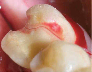 |
| Cracked Tooth Syndrome |
In more recent times the
definition has been amended to include, ‘a fracture plane of unknown depth and direction
passing through tooth structure that, if not already involving, may progress to
communicate with the pulp and or periodontal ligament
In the late 1970s, Maxwell and
Braly advocated the use of the term incomplete tooth fracture. According to
Luebke, fractures are either complete or incomplete, although, other terms such
as
• Incomplete tooth fracture
• Cracked tooth syndrome
• Split tooth syndrome
• Green stick fracture
• Hairline fracture
• Cuspal fracture odontalgia
are also known.
Classification of Cracked Tooth Syndrome
Several classifications have
been proposed based on:
(a) The type or site of the crack,
(b) the direction
and degree of the crack,
(c) the risk of symptoms,
(d) pathological processes.
Cracked teeth can be
classified on the basis of pulpal or periodontal involvement and the extent of
crack.
Class A: Crack involving
enamel and dentin but not pulp.
Class B: Crack involving pulp
but not periodontal apparatus.
Class C: Crack extending to
pulp and involving periodontal apparatus.
Class D: Complete division of
tooth with pulpal and periodontal apparatus involvement.
Class E: Apically induced
fracture
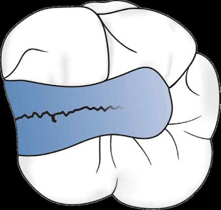
Two classic patterns of crack
formation exist:
1. The first occurs when the crack
is centrally located, and following the dentinal tubules may extend to the
pulp.
2. The second is where the
crack is more peripherally directed and may result in cuspal fracture
Prevalence
1. The condition presents mainly
in patients aged between 30 and 50 years
2. Early epidemiological
survey by Cameron in 1976 seemed to suggest that the condition was much more prevalent
among female dental patients. more recent studies that both sexes seem to be
equally affected.
3. Most cases occur in teeth
with class I restorations (39%) or in those that are unrestored (25%)
4. Mandibular molars (2nd
> 1st molar) are most commonly affected, followed by maxillary
premolars and maxillary molars, while mandibular premolar teeth seem to be
least affected.
5. Lower first molar teeth are
usually the first permanent teeth to erupt into the dental arch, they are most
likely to be affected by the condition of dental caries, followed by the
need of subsequent restorative
intervention. Also wedging effect’ inflicted upon lower first molar teeth from
the prominent mesio-palatal cusp of maxillary first molar teeth may also be
contributory.
6. The transverse ridge of the
maxillary molars may provide structural reinforcement and account for the lower
incidence of fracture in these teeth.
7. According to Homewood,
fractures tend to occur in a direction parallel to the forces on the cuspal
incline.
8. It has also been suggested
that most cracks tend to run vertically (as opposed to horizontally) and usually
run in a mesio-distal direction along the occlusal surface and may involve one
or both of the marginal ridges respectively
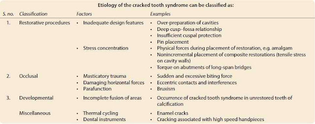
Etiology/Causes of Cracked Tooth Syndrome
1. The aetiology of incomplete
fractures of posterior teeth is multi-factorial. In an article by Guersten et
al.
2. Lynch et al. have
subdivided the causes of cracks into four major causative categories, hence:
‘restorative procedures’, ‘occlusal factors’, ‘developmental conditions’ and
‘miscellaneous factors.
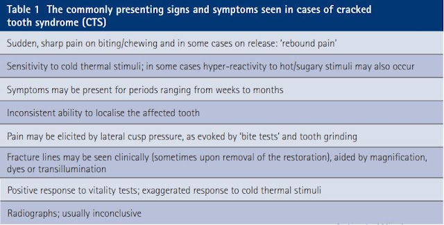
Clinical Symptoms of Cracked Tooth Syndrome
1. Sharp pain when biting, or
when consuming cold food/beverages. Pain increases as the applied occlusal force
is raised
2. Sensitive for years because
of an incomplete fracture of enamel and dentin that produces only mild pain.
3. Other symptoms may include
pain on release of pressure when fibrous foods are eaten, ‘rebound pain’
4. Eventually, this pain
becomes severe when the fracture involves the pulp chamber also. With the
involvement of pulp, Pulpal and Periodontal symptoms may occur.
5. Tooth is sensitive to Cold
and Sweets. The absence of heat induced sensitivity may also be a feature
The type, duration and the
stimuli of pain has important implications for both diagnosis and treatment.
The Commonly Signs and Symptoms are-
The Commonly Signs and Symptoms are-
1. Dentin pain - A brief,
sharp twinge.
2. Pulpal pain - The deep,
demanding, radiating pain precipitated by thermal shock to an inflamed pulp.
The pain at times may be spontaneous.
3. Periodontal pain - The
aggravating throbbing of a sore tooth
Diagnosis
Diagnosing CTS has been a
challenge to dental practitioners and is a source of frustration for both the
dentist and the patient
The ease of diagnosis varies
according to the position and extent of the fracture. Mandibular molars (2nd
> 1st) and maxillary premolars are the most commonly affected
teeth. The tooth often has an extensive intracoronal restoration.
Dental History
History of dental treatment
involving repeated occlusal adjustments or replacement of restorations, which
fail to eliminate symptoms.
The patient will give a
history of pain on biting on a particular tooth, often occurring with foods
that have small, discrete, harder particles in them, for example, bread with
hard seeds or muesli.
Clinical Examination
Includes the presence of facets (identifies
teeth involved in eccentric contact and at risk from damaging lateral forces)
The presence of localized periodontal
defects (found where cracks extend subgingivally),
Symptoms on sweet or thermal
stimuli.
Removing existing restorations
and stains aid in the visualization of the crack.
Visual Examination
Visual inspection of the tooth
is useful, but cracks are not often visible without the aid of magnifying
loupes.
Tactile Examination
one should gently pass the tip
of sharp explorer along the tooth surface, it may catch the crack.
Exploratory Excavation
Aids in Visual Diagnosis .
Consent of the patient should
be taken before excavation since it is not guaranteed that a fracture will be
found underneath any removed restoration.
Removal of existing
restorations may reveal fracture lines.
Percussion Test
They are seldom tender to
percussion (when percussed apically).
Periodontal Probing
With the help of periodontal
probing, one can distinguish between a cracked tooth and a split tooth when the
fracture line extends below the gingiva, thereby causing a localized
periodontal defect.
Careful probing must be
performed to disclose the presence of an isolated periodontal pocket.
However, isolated deep probing
often indicates the presence of split tooth, which predicts a poor prognosis.
Dye Test
Various dyes like Gentian
Violet or methylene blue stains can be used to highlight fracture lines. The
disadvantage of this technique is that it takes at least 2-5 days to be
effective and may require placement of a provisional restoration.
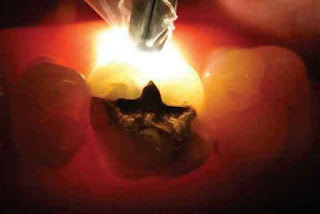
Transillumination
The use of fiberoptic light to
transilluminate a fracture line is also a method of diagnosing cracked tooth
syndrome.
Transillumination is probably
the most common modality for traditional crack diagnosis.
There are two drawbacks to
using transillumination without magnification.
First, transillumination
dramatizes all cracks to the point that craze lines appear as structural
cracks.
Second, subtle color changes
are rendered invisible.
Transillumination with a
fiber-optic light and use of magnification will aid in visualization of a
crack.
Bite Test
Orange wood stick, rubber
wheel or the tooth slooth are commonly used for detection of cracked tooth.
Tooth slooth is small pyramid
shaped, plastic bite block with small concavity at the apex which is placed over
the cusp and patient is asked to bite upon it with moderate pressure and
release.
The pain during biting or
chewing especially upon the release of pressure is classic sign of cracked
tooth syndrome.
Commercially available
diagnostic tools to undertake ‘bite tests’ include products such as ‘Fractfinder’
and ‘Tooth Slooth II’
Radiographs
Radiographs are not of much
help especially, if, crack is mesiodistal in direction. Even the buccolingual
cracks will only appear if there is actual separation of the segments or the
crack happens to coincide with the X-ray beam.
Taking radiographs from more
than one angle can help in locating the crack
Microscopic Detection
Experienced clinicians using a
clinical microscope have reached a general consensus that ×16 provides an ideal
magnification level for the evaluation of enamel cracks, with a range from ×14
to ×18.
Use of the clinical microscope
makes possible the treatment of asymptomatic but structurally unsound posterior
teeth.
Surgical Exposure
If a fracture is suspected, a
full thickness mucoperiosteal flap should be reflected for visual examination
of root surface.
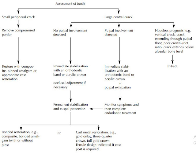
Treatment/Management of Cracked Tooth Syndrome
Urgent care of the cracked
tooth involves the immediate reduction of its occlusal contacts by selective
grinding of tooth at the site of the crack or its antagonist.
Definitive treatment of the
cracked tooth aims to preserve the pulpal vitality by providing full occlusal
coverage for cusp protection.
When crack involves the pulpal
floor, endodontic access is needed but one should not make attempts to chase down
the extent of crack with a bur, because the crack may become invisible long
before it terminates.
Endodontic treatment can
alleviate irreversible pulpal symptoms.
If the crack is partially
visible across the floor of the chamber, the tooth may be bonded with a
temporary crown or orthodontic band. This will aid in determining the prognosis
of the tooth and protect it from further deterioration till the endodontic
therapy is completed.
A decision flowchart of treatment options is presented
A decision flowchart of treatment options is presented
Prevention
• Awareness of the existence
and etiology of cracked tooth syndrome is an essential component of its
prevention.
• Cavities should be prepared
as conservatively as possible.
• Rounded internal line angles
should be preferred to sharp line angles to avoid stress concentration.
• Adequate cuspal protection
should be incorporated in the design of cast restorations.
• Cast restorations should fit
passively to prevent generation of excess hydraulic pressure during placement.
• Pins should be placed in sound
dentine, at an appropriate distance from the enamel to avoid unnecessary stress
concentration.
• The prophylactic removal of
eccentric contacts has been suggested for patients with a history of cracked
tooth syndrome to reduce the risk of crack formation


Very informative... excellent work!!!
ReplyDeleteVery valuable information, it's not like all the blogs that we find here, congratulations, I was looking for something like this and found it here. Thank you for sharing this blog here. Orthodontic Clear Aligner Clinic
ReplyDelete