Cementum
History
Physical Properties of Cementum
- Color- Yellowish in colour and lighter when compared with dentin. It lacks a shiny surface, by which it can be distinguished from enamel.
- Hardness- The hardness is less than that of enamel, dentin and bone.
- Permeability- Cementum is permeable to water and inorganic ions in normal conditions. The permeability decreases with age.
- Thickness- Cementum varies in thickness. It is thinnest near the cervical region (20–50 μm) and gradually becomes thicker towards the apex, where it measures about 150–200 μm
Chemical Properties of Cementum
- Cementum consists of around 45–50% inorganic components and 50–55% organic components and water.
- It consists of calcium and phosphate in the form of hydroxyapatite crystals.
- Fluoride ions are present in higher concentrations in cementum than in any other hard tissue of the body. The magnesium content of cementum is half of that present in dentin.
- The organic portion of cementum mainly consists of type I collagen.
- Collagen types III, V, VI, XII and XIV are also seen in cementum.
Cementoid
The newly deposited cementum is an organic matrix consisting of collagen and ground substance and is called cementoid tissue. This later undergoes mineralization to form cementum
Classification of Cementum
Based on cellularity
(a) Acellular cementum: Cementum without cells
(b) Cellular cementum: Cementum with cells
Based on the time of origin
(a) Primary cementum: It is the first-formed cementum and is acellular. It develops slowly as the tooth erupts, extending from the cervical portion to the coronal two-third of the root. It consists of intrinsic and extrinsic fibres (PDL).
(b) Secondary cementum: It forms after the tooth reaches the occlusal plane. It consists of extrinsic and intrinsic fibres and is present in the apical third and the inter-radicular portions of the teeth.
Based on the presence of collagen fibrils
(a) Afibrillar cementum: Cementum without collagen fibrils
(b) Fibrillar cementum: Cementum with collagen fibrils
Based on the origin of the matrix fibres
(a) Extrinsic cementum: Cementum with matrix fibres derived from the periodontal ligament
(b) Intrinsic cementum: Cementum with matrix fibres derived from cementoblasts
Acellular cementum appears structureless.
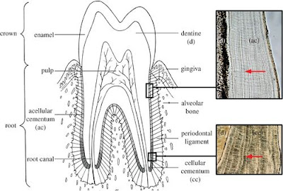
It covers the cervical and middle third of the root. Since it is deposited first, it is also called the primary cementum.
The innermost part of the acellular cementum, near the dentin, will have collagen fibres secreted by the cementoblasts and this part is called the primary acellular intrinsic fibre cementum.
The rest of the primary cementum consists of fibres secreted by periodontal ligament fibroblasts and this portion is called primary acellular extrinsic fibre cementum.
The bulk of the primary acellular cementum is formed by extrinsic fibres because it is secreted after the formation of the periodontal ligament.
Cellular cementum is seen at the apical third of the root.
Since it is deposited after the acellular cementum, it is known as the secondary cementum.
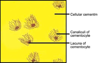
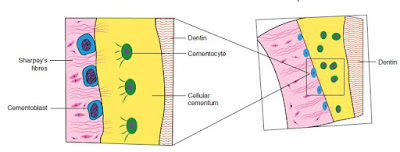
The formation of the cellular cementum is faster, leading to the entrapment of cementoblasts in the matrix.
The cellular cementum is of two types: cellular mixed fibre cementum (consisting of bone, intrinsic fibres and extrinsic fibres) and cellular intrinsic fibre cementum.
A peripheral layer of cementoid with cementoblast lining is always seen in cellular
cementum.
It lacks cells.
The extrinsic fibres are Sharpey’s fibres derived from the periodontal ligament.
It covers the cervical and middle third of the root. Since it is deposited first, it is also called the primary cementum.
The innermost part of the acellular cementum, near the dentin, will have collagen fibres secreted by the cementoblasts and this part is called the primary acellular intrinsic fibre cementum.
The rest of the primary cementum consists of fibres secreted by periodontal ligament fibroblasts and this portion is called primary acellular extrinsic fibre cementum.
The bulk of the primary acellular cementum is formed by extrinsic fibres because it is secreted after the formation of the periodontal ligament.
Cellular Cementum
Since it is deposited after the acellular cementum, it is known as the secondary cementum.
The formation of the cellular cementum is faster, leading to the entrapment of cementoblasts in the matrix.
The cellular cementum is of two types: cellular mixed fibre cementum (consisting of bone, intrinsic fibres and extrinsic fibres) and cellular intrinsic fibre cementum.
A peripheral layer of cementoid with cementoblast lining is always seen in cellular
cementum.
Cementocytes
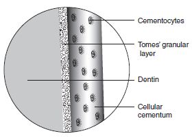
Cementocytes are resting cells seen entrapped in the matrix of cellular cementum. They are similar to osteocytes and lie in a space called lacunae.Acellular Afibrillar Cementum
Acellular afibrillar cementum is seen over the enamel near the cementoenamel junction.Acellular Extrinsic Fibre Cementum
This type of cementum is present in the cervical third and covers two-third of the root of the tooth and is formed in both the pre-eruptive and the post-eruptive phases.It lacks cells.
The extrinsic fibres are Sharpey’s fibres derived from the periodontal ligament.
Cellular Intrinsic Fibre Cementum
Seen in the apical region of the tooth.
The intrinsic fibres are secreted by cementoblasts. It does not possess the ability to serve as anchorage.
Mixed Fibre Cementum
This type of cementum consists of both extrinsic and intrinsic fibres. It is seen in the apical and furcation regions.
Cemento-Enamel Junction
Junction between the cementum and the enamel at the cervical portion of the teeth. Three
types of junctions exist:
Sharp junction (butt joint): The cementum meets the enamel in a sharp line. This type is seen in 30% of teeth.
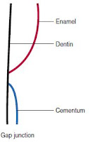
Gap junction: The cementum does not meet the enamel in the cervical region, and there is no CEJ. A portion of the root in the cervical region will be devoid of cementum.
Overlap junction: The cementum overlaps the enamel
CEMENTO-DENTINAL JUNCTION
Cementodentinal junction (CDJ) between cementum and dentin is usually smooth but can be scalloped in deciduous teeth.
RESORPTION AND REPAIR OF CEMENTUM
In cases of excessive trauma, localized areas of resorption occur in cementum. In such conditions, repair takes place by deposition of cementum by cementoblasts.
Causes of Cementum Resorption
There may be local or systemic causes of cemental resorption. These are as follows:
Local Causes
Local conditions which give rise to cemental resorption are as follows:
1. Trauma from occlusion
2. Cysts and tumors
3. Periapical pathology
4. Excessive orthodontic force
5. Embedded teeth
6. Replanted and transplanted teeth.
Systemic Causes
Systemic conditions causing or inducing cemental resorption are as follows:
1. Deficiency of calcium
2. Deficiency of vitamin A and D.
3. Hypothyroidism
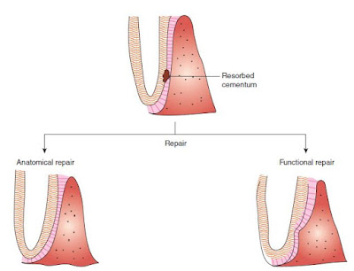
The repair tissue that forms is cellular type. Repair in cementum occurs in two ways:
Anatomical repair: The resorbed area is completely filled up with cementum. The continuity of the root surface and the periodontal attachment is re-established.
Functional repair: The resorbed area is filled up partially, resulting in a bay-like defect on
the root surface. The alveolar bone forms a bony projection to establish the normal physiologic width of the periodontal attachment.
FUNCTIONS OF CEMENTUM
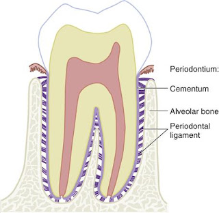
1. Anchorage (attachment): Cementum provides a medium for the attachment of periodontal ligament fibres, thereby helping in binding of tooth with alveolar bone. Acellular cementum is mainly involved in the attachment process.
2. Adaptation: Masticatory forces cause occlusal wearing, thereby reducing the tooth length. By the continuous deposition of cellular cementum at the apical region of root, the decrease in tooth length is compensated and an occlusal relationship is maintained.
3. Repair: Fracture or resorption occurring in the roots is repaired by the deposition of cellular cementum, because it forms at a faster rate.
Incremental Lines of Salter
The rhythmic deposition of cementum is represented by incremental lines of Salter. They do not have a regular rhythmic distribution as seen in other mineralized tissues of tooth structure.
They are unevenly spaced. Incremental lines in acellular cementum are arranged more regularly and evenly than in the cellular cementum.
Bone and Cementum
Phenotypic similarities between the osteoblast forming the bone and the cementoblast forming the cellular intrinsic fibre cementum have been proven. There are few more similarities and differences between the bone and cementum.
Cementum Anomalies
Hypercementosis
Hypercementosis means abnormally prominent thickness of the cementum on root surface. This abnormality may affect all the teeth of dentition together or may occur on a single tooth of dentition.
Local and Generalized Hypercementosis
Localized Hypercementosis is found at a single location anywhere on the root surface. The examples of localized hypercementosis are cemental spikes, and excementosis.
Generalized hypercementosis that involves roots of all the teeth of a dentition is present in certain conditions such as:
i. Paget’s disease of bone [Hereditary]
ii. Chronic periapical infection.
iii. Non-functional teeth without any antagonist.
Difference between Cemental Hyperplasia and Hypertrophy??
Cemental Hyperplasia Hypercementosis is also called as cemental hyperplasia when cemental growth does not help in increasing functioning of the tooth or it occurs in non-functional teeth, like hypercementosis due to periapical infection.
Cemental Hypertrophy If cemental overgrowth improves or helps in the functioning of teeth, this is referred as cemental hypertrophy. Cemental spike generally develops from extensive occlusal forces or tensional orthodontic force. This provides a greater surface area for the attachment of periodontal fibers.
Cementicle
Cementicles are round lamellated cemental bodies that lie free in the periodontal ligament space or are attached to the root surface. Mostly they are found in an aging person along the root. They may be found at the site of trauma.
Cementoma
Cementoma is also called benign cementoblastoma or cemental dysplasia. These are cemental masses situated at the apex of the root. These are included in the category of a slowly growing odontogenic neoplasm and may cause expansion of jaw.
These are actually true neoplasms of functional cementoblasts. These occur more in females than males under the age of 25 years and the mandibular first molar is the most common site of development





Superb explanation
ReplyDelete