Tooth Enamel
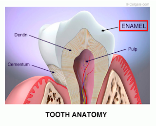
Tooth Enamel is an ectodermal derivative and is the most highly mineralized tissue known.
It is the hardest tissue of the body, which covers entire surface of the anatomical crown of all the teeth.
 |
| Tooth Enamel |
Enamel provides shape and contour to the crown of teeth. It is also known as Substantia adamantia.
1) Physical Properties
A) Thickness- Enamel is thicker in the cusp of the molars and premolars, reaching up to a maximum of 2–2.5 mm, whereas at the cervical region, it almost thins down to a knife edge.
B) Hardness and Density- Varies in different parts of the crown. The hardness and density decreases from the surface of the enamel to the dentinoenamel junction (DEJ) and from the cuspal or incisal region to the cervical margin.
C) Specific Gravity- The specific gravity of enamel is 2.8
D) Color- The colour of the enamel varies from light yellow to greyish white. The colour depends on the translucency of the enamel, which is mostly associated with a variation in the degree of calcification and homogeneity of the enamel. The refractive index of enamel is 1.62.
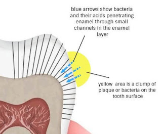
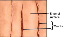
E) Permeability- Enamel acts like a semipermeable membrane, allowing complete or partial passage of certain molecules. The permeability of the enamel is the result of the presence of cracks and microscopic spaces on the surface of enamel which allow the penetration of fluids.
F) Brittle- The structure and hardness of the enamel makes it brittle.
G) Density- The density of enamel is 2.8 to 3 g/ml.
H) KHN- It has a Knoop hardness number (KHN) of 343 while dentin has 68 KHN and cementum has 40 KHN. Enamel resists masticatory impact of about 10 to 20 kg per tooth.
2) Chemical Properties
The enamel is made up of 96% inorganic material and 4% organic matter and water by weight.
The inorganic content is mainly hydroxyapatite crystal and is present 92 to 98 percent by volume of total inorganic matter
The organic matrix of enamel is made up of two types of non-collagenous protein components: amelogenins (90%) and non-amelogenins (10%).
The non- amelogenins constitute the enamel proteins enamelin, ameloblastin, sulphated protein and tuftelin.
- Read About
3) Structure of Enamel
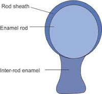
Enamel is composed of the following.A. Enamel rods (prisms)
B. Rod sheaths
C. Inter rod substance (cement)
A. Enamel Rods (Prisms)
An
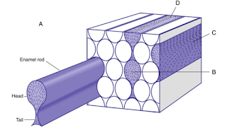
enamel rod is a long, thin structure extending
from the dentino-enamel
junction
to the
Follows
a
tortuous course; thus the length
of an enamel rod may be greater than the
thickness of
enamel.
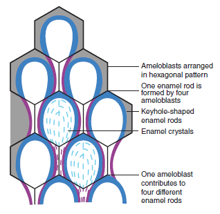
Each rod is formed by four ameloblasts. One ameloblast forms the rod head, a part of two
ameloblasts form the neck, and the tail is formed by a fourth ameloblast. Each ameloblast
contributes to four different rods.
In
a cross-section
of human
enamel, many rods resemble fish-scales.
Direction
of rods In general, the rods are directed at right
angles to
the dentino-enamel
junction
and the tooth surface.
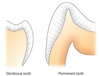
In cervical
regions of deciduous and permanent teeth, the directions of
enamel rods are
different
horizontal.
Near the tip of the cusp and incisal edge, the rods gradually change to increasingly oblique
direction.
Gradually they become nearly vertical at the tip of the cusp
and incisal region.
In the cervical region, the rods run slightly apically from dentin surface to outer enamel
surface
In deciduous teeth: The arrangement of rods in the occlusal two-thirds in deciduous teeth
is similar to that in permanent teeth. At the cervical and central part of the fossa and pit,
they are nearly horizontal.
The diameter of the rod increases from the dentino-enamel junction towards the outer
surface of enamel in a ratio of about 1 : 2. The number of enamel rods has been estimated
as ranging from 5 million in the lower lateral incisor to 12 million in the upper first molar.
The rods are larger at cusp tips and shorter at the cervical region.
The head or body of the enamel rod is the broadest part and is about 5 microns wide, and
the elongated thinner portion called as tail is about 1 micron wide. The rod, including both
head and tail, is about 9 microns long.
In cross-section, the enamel rods appear as round, hexagonal or oval. Recent studies with
electron microscope have shown that a more common pattern of enamel rods in cross
section looks like a keyhole or paddle-shaped prism.
B. Rods Sheath
Under
light microscope, a distinct thin layer is seen
peripheral to
the rods. It has a different
refractive index,
stains darker
and is more acid-resistant than the rod. It is
less calcified
and
contains more organic substance like
enamel protein.
This layer is known as rod sheath.
C. Inter-Rod Substances
Light
microscope revealed that the rods are cemented
together by
inter-rod substance,
which has slightly higher refractive index
than the
rods. The crystals are arranged
in a
different direction in the inter-rod region.
4) Other Structures
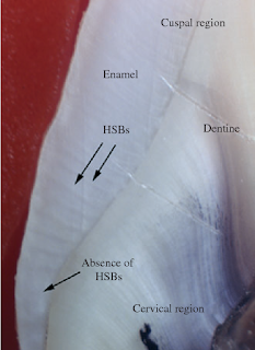
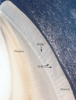
A) Hunter-Schreger Bands
These
are alternating dark and light bands which are best visualized in longitudinal
ground
section under oblique reflected light.
It is produced solely by changes
in the rod direction.
The light bands are referred to as diazones and the dark
bands are called as parazones.
The angle between the diazones and
parazones is
approximately of 40 degrees
B) Incremental Lines of Retzius
These
are rhythmic successive apposition of layers of enamel during formation of the
crown.
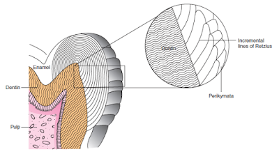
When a ground section of a tooth is seen under a light microscope,
concentric brown lines
are seen in the enamel. These are called incremental
lines of Retzius or Striae of Retzius.
The incremental lines (striae) of
Retzius are more
frequently seen
in permanent teeth and
less frequently in
deciduous teeth
and prenatal enamel.
C) Structureless Outer Enamel Layer
30
microns thick
found most commonly towards the cervical
area and
less often on cusp
tips.
This structure less layer
is called prism
less enamel
and found
in all
deciduous teeth and in
70 percent of the permanent
teeth.
D) Perikymata
wave-like,
transverse grooves. They are shallow furrows
and most probably the external
manifestation of
incremental lines of Retzius. They are
continuous around
a tooth and
usually lie parallel to each
other and
to the cemento-enamel
junction.
E) Enamel Rods Ends
The
enamel rod ends are concave and vary in depth and
shape.
They may contribute to the
adherence of plaque
material with
a resultant caries attack, especially in young
people.
F) Enamel Lamellae
Enamel
lamellae are very thin, leaf-like structures,
sometimes visible
to naked eye. They
extend from the enamel surface
towards the dentino-enamel
junction,
rarely
extending into
dentin. The enamel lamellae contain mostly
organic material.
Enamel lamellae can be differentiated into three types:
Type A lamellae composed of poorly calcified rod segment.
Type B lamellae composed of degenerated cells.
Type C lamellae arising in erupted teeth where the cracks are filled with organic matter and
debris from saliva.
Type A is restricted to enamel and type B and C may reach the dentin
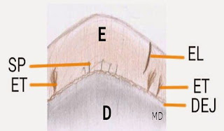
G) Enamel Cracks
Narrow,
fissure-like structures that are present on almost
all surfaces.
They are actually the
outer edges of enamel
lamellae.
They originate from dentino-enamel junction
and
run at
right angles to it.
H) Neonatal Lines
In deciduous teeth, the enamel develops partly before and
partly after
birth. The line or
boundary between the two
portions of
enamel in deciduous teeth is known as
neonatal line
or neonatal ring.
It
appears due to
the abrupt
change in the environment and nutrition of
the
newborn
(infants). It is an accentuated incremental
line of
Retzius.
I) Nasmyth’s Membrane (Primary Enamel Cuticle)
Nonmineralized
usually found
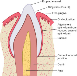
between the epithelium of dentogingival junction
and the
enamel surface. It is formed by an
accumulation of basal lamina material produced by
the
junctional
epithelium of the dentogingival junction.
The final
act of the ameloblast
cell is
secretion of a layer covering the end of the enamel
rod.
J) Pellicle or Salivary Pellicle
After
tooth is cleaned, salivary proteins and glycoproteins
having strong
affinity for enamel
get adsorbed to the
enamel surface
very quickly and form a very thin layer
called the
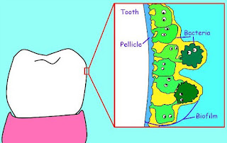
salivary pellicle.
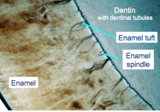
K) Enamel Tufts
Enamel
tufts are hypocalcifed enamel rods and interprismatic substance that originates
at
the dentinoenamel junction and extends into enamel for about one-third to
one-fifth of its
total thickness. They are
known as
enamel tufts because they look like tufts of grass
projecting into
enamel.
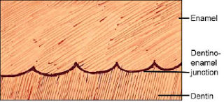
L) Dentino-Enamel Junction
The
dentinoenamel junction is a scalloped interface between
the enamel
and dentin. Dentin
has pitted surface, which
supports the
enamel.
Small
curved
projections of
enamel fit into
small concavities of the
dentin.
M) Enamel Spindle
Odontoblastic
processes sometimes cross the dentinoenamel junction and
get entrapped in
the enamel matrix. Since mostly
they are thickened at their end they have
been termed
as
enamel spindles. They may serve as pain
receptors, thereby explaining the enamel
sensitivity experienced
by some patients during cavity preparation.
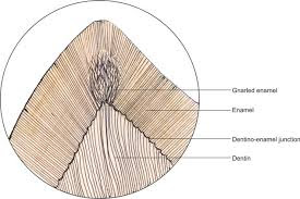
N) Gnarled Enamel
The
enamel rods at the cuspal and incisal region appear
intertwined,
twisted and inter
twisted, and are more
irregular.
Such kind of optical
appearance of
enamel is called as
gnarled enamel. They
are more
so at the cuspal region than incisal region.
O) Enamel Droplets or Enamel Perals
Occasionally,
the cells of the epithelial root sheath remain
adherent to
the dentin surface,
they may differentiate into
functioning ameloblasts
and form small round islands of enamel.
Such droplets of enamel are called enamel
pearls.
They may be found near or in the
bifurcation or
trifurcation of
the roots of permanent molars.
# Want to Solve MCQs on Ename- Click Here





Thank you!
ReplyDeleteyour welcome doc
Delete