Contents
What is Dental Pulp?
The
Pulp is a soft mesenchymal connective tissue that occupies pulp cavity in the
central part of the teeth.
Morphology of the Pulp
The
shape of the dental pulp is similar to that of the
tooth.
It
has coronal and radicular parts. The coronal pulp resembles the
morphology of the crown.
The
extension of the coronal pulp high into the cusps is known as pulp horn.
The
radicular portion of the pulp is seen in
the innermost part of the roots, where it is called the root
canal.
There
are lateral branches to these canals
called the accessory canals.
At
the apical end
of the root canal is the opening called the apical foramen.
Through
the apical foramen, the pulp communicates with the periapical tissues.
The
average size of the apical foramen of maxillary
teeth is 0.4 mm and mandibular teeth is 0.3 mm.
The
volume of the pulp is very little—on an average,
0.02 cc per tooth. The total pulp
volume of all the teeth is 0.38 cc.
- Read About
- Pulp Vitality Tests with Classification and Recent Advances
- Dentin- Microscopic Structure, Properties, Types and Functions
Histology of the Pulp
The
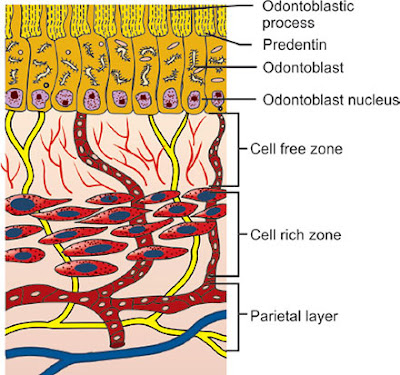
coronal pulp when viewed under the microscope
shows the following zones.
A. Odontoblastic
zone
B. Cell-free
zone of Weil
C. Cell-rich
zone
D. Pulp core
A. ODONTOBLASTIC ZONE
Tall columnar odontoblasts are arranged in Palisading pattern forming a
single layer in the Peripheral area of
the pulp.
Lines the outer pulpal wall and
consists of the cell bodies of odontoblast.
B. CELL FREE ZONE OF WEIL
Cell-free
zone of Weil appears as a space between the
odontoblastic zone and the cell-rich zone.
There
are no cells in this zone. However, there are a few fibres which run through this zone.
C. CELL RICH ZONE
Cell-rich
zone contains numerous cells, fibroblasts being
the highest population.
Undifferentiated mesenchymal
cells, mast cells and macrophages are also seen.
D. PULP CORE
It is central region of the pulp
Contains major blood vessels and nerve of the pulp
Pulpal cells and fibroblasts are also seen
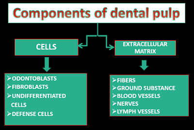
1) Fibroblasts
These
cells form the predominant group
of cells in the pulp.
The
shape of the fibroblasts varies from round to stellate/star shaped.
They
secrete Types
I, III, V and VI collagen and a wide range of
non-collagenous extracellular
matrix components, such as proteoglycans and fibronectin.
They
synthesize and degrade collagen.
2) Odontoblasts
Second most numerous cells in the pulp.
The fully differentiated is a polarized columnar cell with Diameter- 5-7µm, Length- 25-40µm
Constricts to Diameter 3-4µm near pulp, taper to 1µm into dentin
Shape may vary
3) Undifferentiated Cells
These
are totipotent cells which can differentiate into
odontoblasts, fibroblasts or macrophages when
the need arises.
They are found throughout
the cell-rich area and the pulp core
and are present in proximity to blood vessels.
4) Defence Cells
They are histiocytes, macrophages, dendritic
cells, mast cells and plasma cells.
In addition, neutrophils, eosinophils, basophils,
lymphocytes and monocytes are also
seen.
they
respond to situations
that induce inflammation such as dental
caries, mechanical and chemical irritation, trauma
from occlusion, etc
5) Fibers
Collagen
is the major organic component in
the dental pulp
They range in length from
10 to 100 nm and exhibit cross striations at
64 nm
The pulp contains type I and type
III collagens, traces of V and VI are also found.
Type I and III are found in the ratio of 55:45.
Collagen is synthesized and secreted by
odontoblasts and fibroblasts
Odontoblasts secrete
type I and fibroblasts secrete type III.
Elastic
fibres are absent in pulp and is the reason for
dental pulp being termed as specialized connective
tissue.
TYPE I: Present as thick striated
fibrils. Responsible for pulp architecture.
TYPE III : Thinner fibrils, mainly
distributed in cell free and cell rich zone. Contributes to the elasticity of
the pulp.
TYPE
IV : Present along the basement membrane of blood vessels.
TYPE
V & VI : Form a dense meshwork in the form of a thin fibril in the entire
stroma of the pulp.
Collagen fibres arising
from the pulp passing spirally between odontoblasts, enter the predentin
6) Ground Substance
The ground substances
consists of acid mucopolysaccharides and neutral glycoprotein.
These substances are the
environment that promotes life of the cells
Glycosaminoglycans are bulky
molecules and hydrophilic, they form gels that fill most of the extracellular
space
They contribute to the high tissue
fluid pressure of the pulp
What is the pulp richly supplied with?
Blood Supply
Pulp
is a highly vascular tissue
The
velocity of
blood flow and blood pressure is higher when
compared to other tissues
Supplied by the Superior and the
Inferior alveolar arteries
Pulp
is a micro circulatory system which lacks true arteries and veins
The
largest vessels are arterioles & venules which regulate the local
interstitial environment
These arterioles undergo extensive
branching to form ultimately small capillaries
The flow of blood in
arterioles - 0.3
to 1mm/sec
Venules – 0.15mm/sec
Nerve Supply
The
pulp has an abundant nerve supply.
Sensory supply
They
are involved in perception of
pain and transduction
They
are derived from
the branches of the maxillary and mandibular divisions
of the trigeminal nerve
They
give out extensive branches near
the cell-rich zone, forming the parietal
layer of nerves or plexus of
Raschkow
The
plexus contains both large myelinated A-delta
and beta fibres (2–5 μm in diameter) and the
smaller unmyelinated C fibres (0.3–1.2 μm)
Autonomic Sympathetic Nerve Fibres
The sympathetic
nerve fibres originate from the cervical sympathetic
ganglion and join the trigeminal nerve
at its ganglion
They are unmyelinated and follow
the course of the sensory nerves
Regulate
blood flow in the capillary network
by innervation of the smooth muscle cells.
What is the function of pulp in the tooth?
FUNCTION OF THE PULP
Inductive - To induce oral epithelial differentiation into
dental lamina & enamel organ formation.
Formative - Produce the dentin that surrounds
& protects the pulp.
Nutritive - Nourishes the dentin through
odontoblasts & by means of blood vascular system.
Protective -Respond with pain to all stimuli.
Defensive or reparative - Produce reparative
dentin & mineralize any affected dental tubules.
Pulp Stones (Denticles)
Pulp
stones, or denticles are calcified, shiny nodular masses
that occur either single or multiple in numbers within
the coronal and radicular pulp.
The
pulp stones usually
are asymptomatic in nature, but may be symptomatic if the stones impinge on
nerves or blood vessels.
Pulp
stones according to their histological structures, have
been classified into two types– true and false
TRUE DENTICLE
Round or ovoid with
smooth surfaces.
The remnants of HERS
induce differentiation of odontoblasts which form true denticle.
Contain dentinal
tubules and found close to root apex
Rough and have no particular shape
Cause may be degenerating cells,
thrombi and collagen fibrils
Concentric layers of calcified
tissue and usually found in the pulp chamber
Pulp
stones, according to their relation with the dentin of
the tooth, may be divided into three types
Free
Attached
The free pulp stones
are completely surrounded by pulp.
The attached pulp
stones are partly surrounded by pulp and partly fused
with dentin
The embedded pulp stones are completely
surrounded by dentin
Diffuse Calcification
They
are irregular areas of calcification in the pulp tissue that
can be seen as a large mass or fine spicules of calcified tissue.
Appear
as irregular calcific deposits in the pulp.
Pulp
organ may be free of any pathology but may exhibit these changes more
frequently in the roots unlike denticles
Tags:
Dental Histology


The information you have published here is really awesome, as it contains some great knowledge which is very essential for me. Thanks for posting it. childrens dentist near me
ReplyDelete